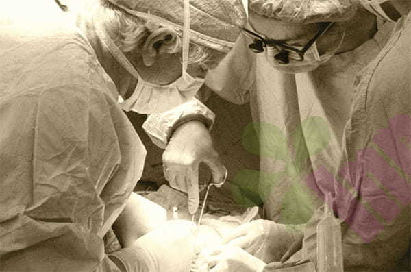
Colostomy skin and mucous membrane separation refer to the separation of the intestinal mucosa and abdominal wall skin at the suture of the colostomy. Colostomy mucocutaneous separation refers to the suture separation between the mucous membrane of the colostomy and the skin of the abdominal wall. After the mucocutaneous breakup of the enterostomy, the open wounds formed can be shallow or deep, usually reaching the subcutaneous layer. It is challenging to collect feces after the separation of the mucous membranes and the skin. The causes include partial necrosis of the intestinal wall mucosa at the open end of the enterostomy, detachment of the mucosal suture, excessive abdominal pressure, wound infection, malnutrition, and diabetes, resulting in poor tissue healing at the stoma mucosal suture, forming an open wound, which requires Regular follow-up and proper nutritional support can play a preventive role.
The principle of dressing selection:
Under sterile conditions, using airtight dressings can keep the wound at a suitable temperature and humidity, which is conducive to forming damaged epithelial cells and promotes the growth of granulation tissue and wound healing. The alginate dressing can be used as a filler in the wound bed at the separation site. It absorbs moisture and forms a gel, which can keep the wound moist and play the role of autolytic debridement. The silver ion dressing can continuously and effectively release silver ions, destroy the bacterial cell membrane, increase its permeability, and lead to the death of bacteria; the silver ion dressing can prevent the formation of bacterial colonies in the wound; destroy the transmission of bacterial substances and cause the bacteria to die. The use of silver ion dressings has both antibacterial properties and promotes wound healing.
Selection and replacement of ostomy bags and accessories:
For patients with significant stoma retraction and skin and mucous membrane separation, a Longterm Medical convex bottom plate, two-piece ostomy bag, and stoma belt are recommended. After the stoma is retracted, it is easy to cause the excrement to accumulate in the depression of the stoma, which is not conducive to the discharge of the waste, and also increases the chance of separation of the skin and mucous membranes, which is not conducive to the healing of the wound. The convex bottom plate can effectively improve the stoma retraction problem, and the convex bottom plate can press the skin around the stoma to make the stoma papilla bulge. After using the convex bottom plate, the stoma nipple is protruded, and the mucous membrane is raised. After the surrounding skin is pressed down, the mucous membrane is even higher than the skin, which improves the retraction phenomenon. Convex baseplates must be used with a suitable ostomy girdle, which increases the pressure on the surrounding skin and allows the baseplate to make complete contact with the skin. The tightness of the belt should be appropriate, and it is fair to be slightly tight so that it does not affect abdominal breathing. The size of the bottom plate ring should be 1-2 mm larger than that of the enterostomy to prevent leakage of excreta to the separation wound. Before attaching the base plate, apply a leak-proof paste around the edge of the stoma and secure it with a belt. Use leak-proof ointment on the distal stoma so that the chassis covers it. For those who have separated the skin and mucous membranes of the stoma, it is recommended to use a two-piece ostomy bag. Compared with the frequent removal of a one-piece ostomy bag, the skin protection effect is better. Try to choose a transparent pack, which can easily observe the stoma bottom plate and whether there is leakage in the separated part of the wound. The ostomy bag can be easily removed and installed, and it is more convenient and straightforward to replace.
Treatment of wounds Nursing measures:
1. The skin around the stoma is cleaned with isotonic saline, debridement, and necrotic tissue removed.
2. The wound at the mucosal separation was filled with alginate dressing to promote the growth of granulation tissue. The outer layer was covered with a dressing, and the skin around the stoma was coated with stoma skin care powder + skin protectant.
3. Paste a plastic leak-proof ring to prevent leakage.
4. Replace the convex chassis and stick the ostomy pocket.
5. Nutritional support
6. If the patient is obstructed and cannot eat orally, they can be given intravenous hypernutrient therapy as prescribed by the doctor. If the patient can eat orally, instruct the patient to eat more high-protein foods, drink more water, and eat less crude fiber foods and foods that are likely to cause stoma obstruction.
7. Anal expansion, follow-up
8. Since mucosal separation may lead to stoma stenosis, patients should be instructed to properly dilate the anus 1-2 times a day for 5 minutes each time. In addition, inform the patient to return to the doctor regularly, and if there is any discomfort, they should seek medical attention at any time.
9. Psychological support, comfort patients, help patients build confidence, and face the stoma correctly.
Prevention Advocacy:
Prevention is better than cure. Evaluate the nutritional status of patients who have just completed stoma surgery, strengthen nutrition, prevent the separation of stoma skin and mucous membranes due to low food, correct hypoalbuminemia in time, and use abdominal girdle in time after surgery to reduce abdominal incisions and the tension around the stoma. When replacing the ostomy bag, pay attention to observing the stoma, whether the adhesion of the bowel and the skin is normal, and whether there is a separation of the skin and mucous membranes of the enterostomy. And control.
For more information on Innomed® dressings, refer to the previous articles. If you have customized needs, you are welcome to contact us; we will serve you wholeheartedly.
At Longterm Medical, we transform this data by innovating and developing products that make life easier for those who need loving care.
References:
Yu Dehong. Enterostomy in the treatment of EM]. Beijing: People's Health Publishing House, 2004: 194.
Dealey C. Thecareofwounds[M]. 2nd Edition. London: MPG
Books Ltd, 1999: 78
Editor: kiki Jia
Date: June 30, 2022

 English
 عربى
عربى Español
 русский
 中文简体
中文简体


